Monday, August 6, 2012

Ankle Sprains

What is an ankle sprain? 
An ankle sprain is when an injury occurs to one or more of the ligaments of the ankle.  The ankle joint is comprised of three bones - the tibia, fibula, and talus.  The tibia is the largest bone of these.  All of these bones are connected by a complex arrangement of ligaments which provide inherent stability and allow this joint to function normally.  An ankle sprain can range from a simple overload of the ligament that heals within several days to the complete rupture of multiple ligaments and subsequent dislocation or subluxation of the ankle.


The severity of an ankle sprain is dependent upon whether the ligament is stretched, partially torn, or  completely torn, as well as the number and location of the ligaments that are affected. 
The most common ligaments ruptured are the anterior talofibular ligament (ATF) and calcaneofibular ligament (CF).


Etiology:

An ankle sprain is generally the result of a twisting type of motion to the ankle joint, or a direct blow that forces the ankle out of its normal anatomic alignment.  Ankle sprains can occur while doing virtually any activity although they are most common while playing sports or while wearing inappropriate shoes. 


Symptoms of an ankle sprain:
Symptoms usually include pain, swelling, and bruising.  Often times the ankle joint will become stiff from all the swelling and inflammation (the body's attempt to immobilize itself).  Also, pain deep within the ankle joint may present.  Walking may be difficult or impossible depending upon the severity of the sprain. 

Pain and swelling may be absent in people who have had previous ankle sprains.  The ankle may feel unstable and "floppy" when walking.  An ankle sprain always requires treatment.  If left untreated, the ankle may develop chronic ankle instability - a condition with persistent discomfort and a continual "giving way" of the ankle.  Additionally, a sprain may be accompanied by a more severe ankle injury such as an ankle fracture, or an osteochondral defect (damage to the cartilage within the ankle joint.)
 

Treatment and Rehabilitation:

Immobilization is generally recommended until pain has subsided enough to begin physical therapy.  Therapy is started as soon as possible to promote healing and increase your range of motion.  Anti-inflammatory medications such as ibuprofen or naproxen are generally recommended initially.  Ice and compression are also used in the initial inflammatory stages of rehabilitation. 

Ankle sprain grading and treatment
Grade 1 - stretched ligament = lace up ankle brace
Grade 2 - partially torn ligament= aircast, transition to lace up ankle brace
Grade 3 - complete tear of ligament = CAM Walker until pain subsides, then aircast with PT, Lace up brace once back to activity. 


Once you return to your sport or full activity it may still be recommended to utilize an ankle brace for stability, and to minimize chances for recurrence. This is generally recommended for 3 months with a complete tear doing normal day to day activities to prevent recurrence.  With sports, the lace up brace should be utilized for 6 months to prevent recurrence. 

Occasionally surgery is indicated.  Usually this is for the person who experiences subjective chronic instability of the ankle from multiple sprains or for sprains that did not heal as expected.  Additionally, cartilage damage within the ankle joint (an osteochondral defect) may need surgery.   
 
Initial Treatment:
This injury should be stabilized as soon as possible either by the emergency room or by a foot and ankle specialist.  Until you are able to be examined by a doctor, the “R.I.C.E.” method should be followed. This involves: 

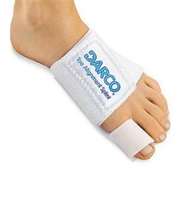
  • Rest. It is crucial to stay off the injured foot, since walking can cause further damage. Non-weightbearing with crutches or a walker is ideal. Generally, an ankle brace will be recommended to stabilize the ligaments initially (regardless of if you are able to bear weight), and then to be used while performing physical therapy. 
  • Ice. To reduce swelling and pain, apply a bag of ice over a thin towel to the affected area for 20 minutes of each waking hour. Do not put ice directly against the skin.
  • Compression. Wrap the ankle in an elastic bandage or wear a compression stocking to prevent further swelling. An ACE bandage is often recommended as initial treatment.
  • Elevation. Keep the foot elevated to reduce the swelling. It should be even with or slightly above the hip level.

Ankle pain and Arthroscopy

Ankle Pain:
 As a brief introduction,: The Ankle Joint (Talocrural Joint) is made up of 3 bones.  The Tibia and Fibula make up the lower leg and these bones are attached to the Talus by a series of ligaments.  Tendons are found all around the ankle joint and cause the foot to go up and down.  This is the action of the ankle joint: up (dorsiflexion), and down (plantarflexion). The up and down motion is what we mean when we say ankle pain.  There may also be pain with straight axial pressure, one may experience a "giving away" type feeling while standing or walking.


The ankle joint rarely sees arthritic changes from wear and tear like the knees and hips do.  Most often ankle arthritis will be a result of some traumatic injury remotely or acutely.  Arthritis is when the cartilage wears away and there is bone on bone.  Along with arthritis comes inflammation, and excess bone growth (bone spurs). 

Ankle fractures and ankle sprains are some of the most common causes of ankle pain and later, arthritis.  Ankle sprains (partial or complete), may have residual pain even months after the injury.  When a ligament is torn, there will be some bleeding into the joint which will later turn into scar tissue and usually have a chronic inflammatory component to it.  Often cortisone injections will alleviate some of the pain associated with inflammation and may help to break up some of the scar tissue.  If several injections have not provided adequate relief of symptoms, an MRI may be indicated to look for more significant causes of ankle pain.
OCD lesions (Osteochondral Defects) may be seen.  These are local areas of cartilage damage or arthritis.     


Osteochondral defects will often present with significant pain and disability.  Occasionally immobilization and rest will provide some relief of the symptoms associated with this pathology.  Often, surgical intervention will be needed to provide long lasting pain relief.  Generally the size of the lesion is predictive of how well the surgery will help to alleviate the pain.  The larger the lesion, the less effective the surgery.  An MRI will often give some idea of the size of the lesion but it may be larger in reality once the joint is visualized arthroscopically (microscopic camera that is inserted into the joint).

Lesions less than 10-12mm in diameter will usually do quite well after arthroscopic surgery.  You may be told to rest for a week or two after this surgery (no weight bearing).  For larger lesions, the surgery is less predictable and requires a longer period of rest (2-4 weeks non weight bearing).  During the surgery, the loose cartilage is debrided and small drill holes are placed in the bone beneath the cartilage.  This stimulates the body to produce fibrocartilage, which is similar to normal cartilage (hyaline cartilage).


Bunions, hammertoes, and neuromas are three of the most common foot issues.  Bunions, also known as hallux valgus, is more frequently seen in women than men but common for both.  Shoes that are ill-fitting generally play a role in making this issue worse but do not usually cause it.  Usually bunions are inherited.  Often a muscle imbalance will increase over time and the deformity may change even with appropriate shoes.  Appropriate shoes have a wide toe-box, adequate length (approx. a thumb thickness between the tip of the great toe and the end of the shoe), and are deep enough shoe to accommodate for any toe deformities.


In addition to wearing different shoes, what else can be done for bunions?
Conservative treatment for bunions is limited but there are several options that can be somewhat helpful.  Bunion splints can help to keep the great toe apart from the second toe and can slow the need for surgery.  Splints can also be used to keep an elevated second toe down.  The Darco splint is one that can help with both of these issues. Wearing this splint at night or while sitting around may provide enough relief to slow the need for surgery.  A cortisone injection into the joint may provide relief if there is an inflammatory component to the bunion or some arthritis.  Additionally, a topical anti-inflammatory patch or gel may provide some relief.  These include Flector Patch, or Voltaren gel.  Conservative treatment usually only provides relief for a short period of time, but is certainly worth trying prior to surgical intervention.


How much does bunion surgery hurt?
The amount of pain that one will have from bunion surgery is highly variable, but it is generally painful since bone work is often performed.  The medial bump is usually cut or chiseled away, and the metatarsal bone is usually cut so as to reorient the great toe joint.  At times, the proximal phalanx of the great toe is cut as well.  Bone cuts are usually stabilized with screws or wires.

In addition to the bone work that is done, there are many other things that factor into the amount of pain one will experience.  Some people are naturally more sensitive to any type of stimulus which will make this surgery more challenging.  Also, individuals taking pain medicine, anti-psychotics/anti-depressants, and sleeping pills will most likely need much stronger medicine after surgery and may benefit from a pain pump where local anesthesia is continually infiltrated around a large nerve for 3-7 days after surgery by a very thin catheter.


How long does it take to heal?
Generally the skin incision will heal in about 10-14 days, and the bone is healed in 6 weeks.  Most of the time you can walk immediately after bunion surgery in a bandage and surgical shoe.  The surgical shoe is usually used for 2 weeks until the bandage is no longer needed.  After 2 weeks you can usually return to a sneaker or running shoe.  After 6 weeks full excercise is generally allowed.  Full exercise means starting with a very low impact activity like a stationary bicycle, then progressing to more traumatic activity as tolerated.  Running and jumping should be the last activities attempted.  You need to increase activity in a stepwise fashion!

The soft tissue will take 3months to a year to loosen up.  The great toe joint will be stiff for quite some time.  If the incision is made dorsally (on the top of the joint), you will often have difficulty flexing the great toe down for some time or perhaps forever.  With a medial based incision, you will not have this difficulty and the scar will not be as visible.  You may need two separate incisions if it is done this way.  The second incision would be made in the webspace between the first and second toes to release a tendon which may be necessary depending on the deformity.

What are the most common complications?
Bunion surgery usually has a fairly high success rate but can be quite painful as mentioned above.  Infections can occur as with any surgery... frequently a pre-operative antibiotic is administered.  Wound healing issues can occur, especially in smokers. Smoking affects all small blood vessels which are critical for healing.  Not only will the skin and soft tissues have a much longer time healing or may not heal, the bone work will take 50-100% longer to heal than in a non-smoker.  The bone work may even go on to a non-union (where it never bonds back together as it is supposed to.). If you smoke, you should strongly consider stopping completely at least 14-23 days before the surgery to prevent these complications, or even not have the surgery.  A smoker who undergoes surgery is set up for failure or revision surgery.

There are specific post-op instructions which need to be adhered to in order to obtain the best result.  There will be restrictions in the type of foot wear needed (post-op shoe, sneakers, etc.), the amount of activity you are allowed, bathing.  These instructions are given so that the most predictable and best outcome is obtained.  If you deviate from these, healing and positional issues will occur that will give you less than a desired outcome or even beget revision surgery.

Often the surgical site will be stiff for an extensive amount of time, up to 6-12 months.  Full muscular strength after surgery usually takes 3-6 months. Although you are often able to bear weight immediately and results are predictable, there is a significant healing phase and you must be patient. The surgery usually takes 45-60 minutes but the healing is the difficult part.


Thursday, April 12, 2012

Heel Pain/Plantar Fasciitis

What is Plantar Fasciitis?

Plantar fasciitis is a painful inflammation of the bottom of the foot between the ball of the foot and the heel.  Usually it is painful first thing after getting out of bed in the morning or after sitting for a prolonged period of time.  This is called "Post-static dyskinesia" - which means pain after being static or immobile for an extended period of time. 

Etiology:

There are several common causes for plantar fasciitis.  These include wearing high heeled shoes or boots frequently, gaining weight, or increased activity level such as walking, standing for long periods of time, or stair-climbing.   

High heeled shoes and boot use can cause this issue by the mere fact that they allow the achilles tendon and fascia to contract and shorten over time.  When you return to normal shoes or barefoot, these structures are tight and put an increased amount of stress on the heel bone (calcaneus).  The pain comes from this pulling on the bone and microtrauma where these structures attach to the bone, and the resultant inflammation and associated pain. 

Gaining weight also will cause a generalized overload to this area and result in microtrauma and inflammation.  Additionally, weight gain can place you at risk for damage to the fat pad which is normally present under the heel.  With additional weight, fat pad breakdown will be an additioal source of pain in addition to the pain of the microtrauma to the plantar fascia.

Recreational activities may play a role in plantar fasciitis even if you are not doing activities that are that different than those you did in the past.  Perhaps a slight change in your walking or running regimen has caused an increased amount of stress on this area of the body.  Sometimes a change in terrain (ie walking on the beach instead of the sidewalk) may be causative.  For people who run, running on concrete sidewalks is more traumatic to the body than runing on asphalt (the concrete is harder and has less give to it.) Additionally shoes that wear out will subsequently place additional stress on different areas of the foot and ankle. 
Symptoms:The main symptom of plantar fasciitis is pain when you walk.  You may also feel pain when you stand and even when you are at rest.  Pain is typically seen as you first step out of bed in the morning or after you get up from sitting for a prolonged period of time.  This occurs because the fibers that had begun to heal in a contracted or shortened position are stretching and tearing.  The pain usually eases up with walking but will often present again after a period of rest or non-weight bearing. 
Examination:
Clinical exam often reveals pain beneath the calcaneus (heel bone).  Pain from side to side is a less common finding.  Generally, plain film xrays will be taken to rule out other less common causes of heel pain such as stress fractures (hairline), calcific tendonosis, fractures, or rarely bone tumors.  Some tests may be performed to examine for nerve issues that can present in the same location or in a location very close to the plantar fascia.  Additionally, MRI may be indicated in certain cases. 
Treatment:
There are two main forms of treatment when discussing plantar fasciitis.  The first form is to control the initial pain, inflammation, and symptoms.  The second form of treatment is aimed at controlling the cause and thus preventing the problem from occurring again.

Conservative measures which target this inital pain, inflammation, and symptoms include anti-inflammatory medications and cortico-steroid injections if pain is severe enough.  Various athletic-type strapping can be applied to the foot and ankle to support the arch and limit overload to the fascia.  Oftentimes, if strapping provides good relief, this will give a decent indication of how much a custom molded orthotic may help.  Additionally, physical therapy is often recommended for various stretching excercises and for application of electrical stimulation and ultrasound treatments. 


Home stretching excercises
which target a tight calf muscle complex will often help out a great deal.  These should be performed 3-4 times daily  for maximum relief


Additionally, certain products can help with stretching such as the Prostretch pictured here.  This device is designed to provide a controlled stretch that is reproducible each time.  This specific device is often used by athletes and by physical therapy departments.

To read more about Prostretch, click here.


The second arm of treatment is aimed at
controlling the cause and preventing a recurrence. 

Often times the foot needs to be supported and there are multiple prefabricated orthotics that can support the foot shape enough to eliminate this pain.  These are much more effective than just simple cushioning pads that are often found at a drug store.  Below are a few samples:

  Orthofeet BioSole-Gel Self Forming


  Powerstep Pinnacle Insoles (Orthotic) Arch Supports


If prefabricated orthotics do not provide enough relief, custom molded orthotics are often recommended.  Custom molded orthotics are specific for each individual foot shape and type.  These devices are created in a lab from a mold or a digital scan.  In contrast to prefabricated orthotics which have a limited lifespan, custom orthotics may last 15-20 years depending upon the materials they are constructed out of.  Generally a firm control layer is the foundation for these devices, and this part holds up for many years.  The top-cover(the part that is against your foot) and the associated cushioning will need to be refurbished from time to time.  For long term control and treatment, custom molded orthotics will provide the best relief.  Over time, the foot may change shape and a new orthotic will need to be fabricated. 

For long-standing or severe plantar fasciitis, a night splint may be recommended.  This is worn at night and when at rest (ie. watching TV with your leg up on a pillow).  The purpose of this splint is to prevent the fascia and calf muscles from contracting while you are at rest.  The fascia begins to heal every time you rest and this helps to allow it to heal in a more stretched out position.
 

Active Ankle Dorsal Night Splint


Additionally, a CAM walker (Controlled Ankle Motion), also known as a walking boot, may be recommended to partially immobilize and allow the body to heal on its own.  This is usually reserved for the most resistant cases of plantar fasciitis.  If utilizing this in a weight bearing fashion does not alleviate the associated pain within 6-8 weeks, you may be instructed to utilize this device with crutches for complete non-weight bearing. Ossur Equalizer Premium Walker - High Top Walker

A below knee cast with crutches is also a viable option to consider prior to any surgical intervention.  This treatment would keep all weight off of this part of the body for a period of 6 weeks or more in an attempt to allow the body to heal on its own. 

Surgical Intervention:
Once conservative measures have been exhausted, surgical treatment may be recommended.  Open surgical procedures are available where both the fascia is released and the spur is can be removed.  However, the spur is usually not causing the pain associated with this procedure, this is found because the body reacts to stress by building more bone. 

Other surgical intervention includes the endoscopic plantar fasciotomy in which two small incisions are made, one on each side of the heel.  A microscopic camera is then inserted and the medial 1/2 of the fascia is released with an endoscopic scalpal.  This procedure usually has very little down time and you will be walking that day after surgery.  

Sunday, April 8, 2012

Acute Calf Pain

Calf pain is often seen after certain activities such as tennis or other raquet sports.  This is frequently called "Tennis Leg." Generally this occurs to the medial head of the Gastrocnemius Muscle.  One will feel a pop or tear at the junction of this muscle and the underlying Soleus Muscle.  This is usually a partial tear of this junction.  Calf pain associated with activity is almost always a soft tissue injury only. 

Other causes of calf pain can include direct trauma which can result in hematoma (blood collection under the skin) or seroma (clearish fluid collecting between muscle bellies or under the skin).  Also, blood clots can occur but rarely from trauma.  Blood clots, also known as DVT (Deep Vein Thrombosis) are addressed in another blog.

Tennis leg is generally diagnosed by clinical exam and history.  Xrays or MRI are rarely indicated.  I may obtain an MRI if the pain is not much better within 2 weeks.  Usually an ACE bandage and rest is indicated for the first 2 weeks after this injury. Crutches may be necessary if the pain is significant. Within 2 weeks the tear usually heals enough to return to gentle activity.  Physical therapy is often started at this time.

Saturday, March 31, 2012

Achilles Tendon Repair Techniques

How do I know if I have sustained and achilles tendon rupture?
The Achilles Tendon is one of the largest and strongest tendons in the body.  It connects the back of the heel bone (Calcaneus) to the calf muscle (Gastrocnemius and Soleus).  This tendon allows running/jumping/walking/lifting to occur normally where the foot is actively plantarflexed.  Normally this tendon allows one to go up on the front part of the foot.  Without this tendon, you are walking on flat foot that does not actively propel very well.

Generally an Achilles rupture is from a traumatic event. Either sports related, work related, or just normal daily activity.  Often doctors will hear a patient comment, "it felt like some one kicked me in the back of the leg. I went to get up and could not use my foot!" Or you may feel like you had a loud "pop" in the back of the leg.  The test for an Achilles Tendon rupture involves squeezing the calf muscle and looking for the foot flexing with this motion.  If the tendon is intact, this will occur easilly.  If it is ruptured, no motion occurs.  Occasionally one needs to compare the affected leg to the healthy one.  An obvious defect is often seen or felt.  In questionable cases, or cases which require additional surgical planning, an MRI may be indicated.

                                                  An obvious defect on the closer leg


Do I need surgery, or is a cast/walking boot enough?
If there is an obvious 1cm gap or greater, surgery is generally recommended.  If the gap is able to be brought together with the foot flexed plantarly, a cast may suffice.  Cast immobilization would generally be indicated in someone who was mostly sedentary or who was bed bound.  For individuals who are fairly active, and who are appropriate surgical candidates, a surgical repair is indicated in almost every case.  The risk of re-rupture is higher if you do not have a surgical repair, and you will need to be immobilized for 6-8, and will not be able to put any weight on your foot.

With surgery, passive motion can be started at 1-2 weeks after repair.  This is assuming you are healing ideally.  Stitches can generally come out at about 2 weeks and you can return to showering. at that time. At 3 weeks after surgical repair, the tendon is at about 50% strength or greater.  It could be argued that with surgical repair, the tendon is much stronger than this due to the strength of the sutures themselves.  Surgery should be considered carefully, as there are many risks with Achilles Tendon Surgery.  But....if you are a person who is fairly active, a surgical repair would usually be recommended.

What are common complications?
In the healthy patient, a full recovery can usually be expected with the appropriate surgical repair. Stiffness and limited range of motion should be expected for several months at least.  The surgical repair can be felt through the skin at times.  In this location, there is not as good of blood flow and skin healing issues are more likely than many other areas of the body.  Again, the most likely people to have post surgical issues are smokers, diabetics, immunocompromised, or the elderly. Infections can occur as with any surgery - they are more difficult to fight in this area because of the decreased blood supply here. Adhesions can occur between the tendon and skin at times as well and the foot will not move as well.

My surgical technique:
An incision is made over the tendon and the paratenon is identified and preserved.  This is a thin layer over the tendon similar to a tendon sheath that allows the tendon to glide smoothly beneath the skin.  It is important to repair this layer cautiously so that the tendon will function optimally. Once the tendon rupture is identified, the ends are freshened up a bit and then re-approximated if possible.


A suture technique called a modified Krackow "Giftbox" is used on both sides of the rupture and then these are tied together.  The knots are tied at a slight distance away from the rupture in a manner that dramatically improves the achilles repair strength over a standard Krackaw repair (1).  Below is a drawing depicting the specifics of this repair technique.  The knots are then buried within the body of the tendon and provide much less iritation through the skin.  This technique has shown to withstand up to 168N of force before failure, where the standard Krackow technique fails at 81N. This technique helps to minimize gap formation and improve the strength of the repair.

 Traditional Krackow Achilles Repair


Modified Krackow "Giftbox" Achilles Repair
(Suture knots tied away from the gap, dramatically
improving strength with less skin irritation)


Additionally, the repair is augmented by an epitendinous cross stitch weave (2) which makes it even stronger and makes the tendon smoother.  The tendon is able to withstand loads sooner and glides much better by utilizing this specific technique.  This has been shown to increase force to failure by 65%. See the drawing and clinical photo below.  I have been using this technique for several years now and have been seeing great results.  Increased strength of the repair which potentially allows patients to return to activity faster with less down time. 


Epitendinous Cross Stitch Weave


1.  Labib,S.A. et al. The "Giftbox" Repair of the Achilles Tendon: A Modification of the Krackow Technique. Foot and Ankle International. 2009.

2.  Lee, S.J. et al. Optimizing Achilles Tendon Repair: Effect of Epitendinous Suture Augmentation on the Strength of Achilles Tendon Repairs. Foot and Ankle International. 2008.